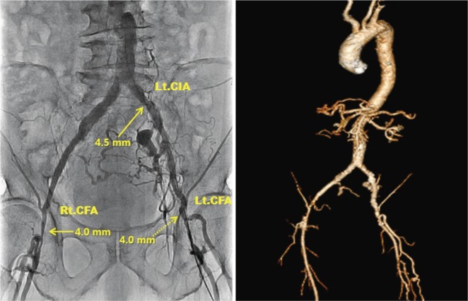
Fig. 2.
Angiography and computed tomography image showed iliofemoral arteries with severe peripheral arterial occlusive disease. Previous stent at Lt. common iliac artery was patent but showed a minimum diameter of 4.5 mm and both common femoral arteries showed a minimum diameter of 4.0 mm. Lt. CIA: left common iliac artery, Rt. CFA: right common femoral artery, Lt. CFA: left common femoral artery.

