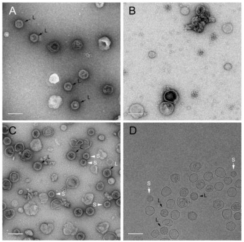
Figure 5.

EM of particles formed upon co-expression of 80α and SaPI1 proteins in S. aureus. (A) 80α gp47 + SaPI1 CpmB (strain ST112); (B) gp47 + SaPI1 CpmA and CpmB (ST114); (C) gp46+gp47+CpmA+CpmB (ST118). (D) Cryo-EM of ST 118. (B) is a P2 pellet; others are sucrose-purified fractions. Examples of small and large procapsids are indicated by S (white arrows) and L (black arrows), respectively. Scale bars equal 100nm.
