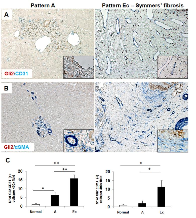
Figure 5. Activated Sinusoidal Endothelial cells and Myofibroblasts are Hedgehog responsive in schistosomiasis mansoni.
Double immunohistochemistry for Gli2 (brown) and CD31 (blue) (A) and for Gli2 (brown) and αSMA (blue) (B) in representative liver sections from subjects with pattern A (left) or with pattern Ec fibrosis (middle). Final magnification ×400, insert ×600. C) Quantification of the immunostaining for Gli2/CD31 (left) and for Gli2/αSMA (right) from selected patients. (Normal=2 subjects, Pattern A=2, Ec=8 (Gli2/CD31) or 5 (Gli2/αSMA)). Results were normalized to the number of double positive cells in normal individuals; means ± S.E.M. are displayed. *p<0.05; **p<0.001.

