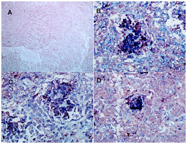
Figure 1. TB patients carrying the two-locus genotype -2518 MCP-1 GG -1607 MMP-1 2G/2G express MCP-1, MMP-1, and MMP9.
We used double IHC analysis of MCP-1 or MMP-1 or MMP-9 and CD68 in paraffin-embedded diseased lung from six TB patients who underwent surgery to remove damaged tissue. We are showing representative data. A: negative control (irrelevant antibodies isotype control); B: MCP-1 (blue) and CD68 (red); C: MMP-1 (blue) and CD68 (light red); D: MMP-9 (blue) and CD68 (light red). Images were acquired at 200X total magnification. IHC analysis shows granulomas with CD68-positive cells (macrophages) expressing copious amounts of MCP-1, MMP-1, and MMP-9.

