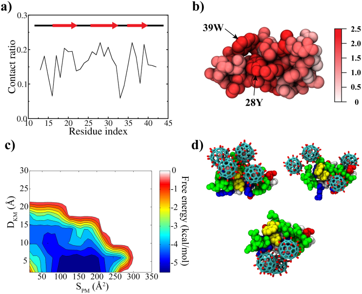
Figure 2.
(a) Site-specific contact ratio of WW domain, where the contact ratio of a residue was obtained by counting the number of frames of the residue in contact with Gd@C82(OH)22 over all time frames; (b) Residues involved in contact with Gd@C82(OH)22; (c) The binding free energy surface, where DKM is the minimum distance between Gd@C82(OH)22 and the signature residues (Y28 and W39) of the WW domain and SPM is the contacting surface area; and (d) Representative binding modes found in the global minimum. Yellow: key residues, white: hydrophobic, green: non-charged polar, red: negatively charged and blue: positively charged residues.

