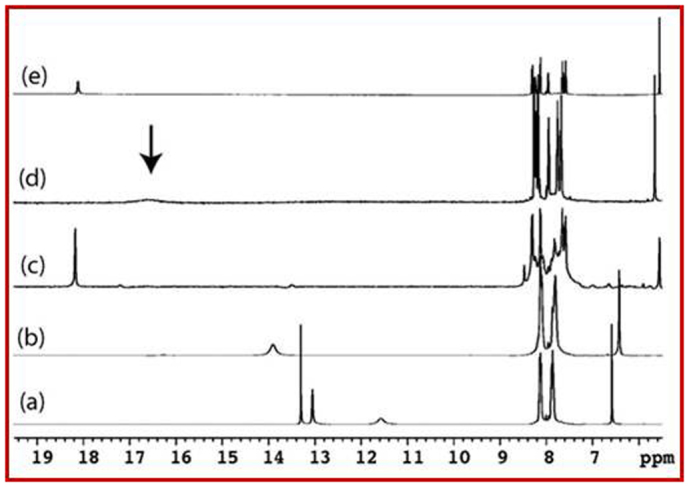
Figure 3. The expanded 1H-NMR spectrum recorded in DMSO-d6 at 500 MHz at 25°C.

(a) Pristine purpurin; (b) Lithiated purpurin electrode [ELP-1] (Cells were discharged (lithiated) from open circuit voltage to 1.5 V vs Li/Li+ in 1 M solution of LiPF6 in 1:1 (v/v) mixture of ethylene carbonate (EC) and dimethyl carbonate (DMC) as electrolyte); (c) Lithiated purpurin electrode [ELP-2] (Cells were discharged (lithiated) from open circuit voltage to 0.02 V in 1 M solution of LiPF6 in 1:1 (v/v) mixture of EC and DMC as electrolyte) and (d) CLP using LiOAc as Li+ source (1:1) and (e) CLP using LiOAc as Li+ source (1:2).
