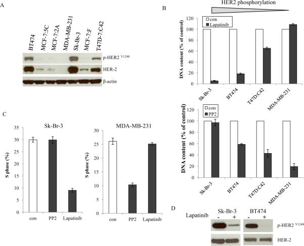
Figure 5. Activation status of HER2 determined the inhibitory effects of the c-Src inhibitor.
5A. Baseline HER2 phosphorylation in different cell lines. Cell lysates were harvested from different cells. Phosphorylated HER2 and total HER2 were examined by immunoblotting with primary antibodies. Immunoblotting for β-actin was determined for loading control. 5B. Inhibitory effects of the HER2 inhibitor and the c-Src inhibitor on cells with elevated HER2 phosphorylation. Sk-Br-3, BT474, T47D:C42, and MDA-MB-231 cells were seeded in 24-well plates in triplicate. After one day, the cells were treated with vehicle (0.1%DMSO), lapatinib (1μM), and PP2 (5μM) in 10% SFS medium. The cells were harvested after 7 days treatment and total DNA was determined as above. 5C. S phase changes after lapatinib and PP2 treatment. Sk-Br-3 and MDA-MB-231 cells were treated with vehicle (0.1% DMSO), lapatinib (1μM), and PP2 (5μM) for 24h. Cells were harvested and fixed with 75% EtOH. Cell cycles were analyzed through flow cytometery. 5D. Blocking HER2 phosphorylation after lapatinib treatment. Sk-Br-3 and BT474 cells were treated with vehicle (0.1%DMSO) and lapatinib (1μM) for 24h. HER2 phosphorylation was examined by immunoblotting with primary antibody. Immunoblotting for total HER2 was determined for loading control.

