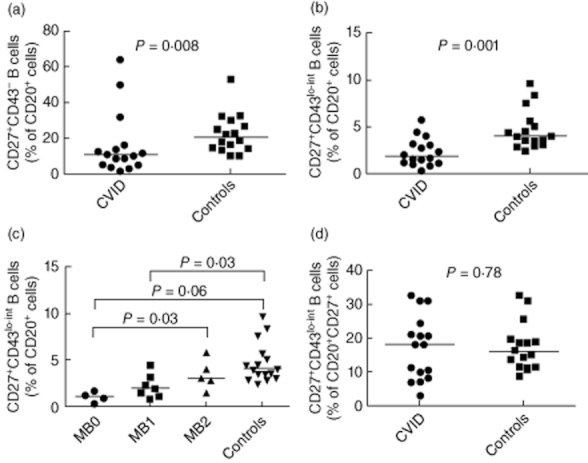
Fig. 5.

Percentages of CD20+CD27+CD43lo–int cells in common variable immunodeficiency (CVID) patients. Whole blood was stained as stated in the methods. Cells were gated as described in Fig. 2. Percentage of CD27+CD43– cells (a) and CD27+CD43lo–int cells (b) within the CD20+ population were measured in CVID patients (n = 16) and healthy controls (n = 33). (c) Percentage of CD27+CD43lo–int cells within CD20+ cells in CVID patients when stratified into respective Piqueras classifications. (d) Percentage of CD27+CD43lo–int cells within all CD27+ cells in CVID patients and healthy controls.
