
Fig. 3.
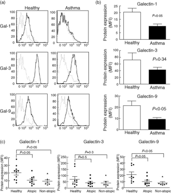
Surface expression of galectin (gal)-1 and gal-9 is reduced in leucocytes from induced sputum of asthma patients. (a) Cells from sputum samples were stained as in Fig. 2 and galectin expression was analysed on macrophages (CD16+ HLA-DR+). Representative histograms from a healthy donor and an asthma patient are shown. Isotype control (dotted line), gal expression (solid line). (b) Gal-1, gal-3 and gal-9 expression on leucocytes from asthma (n = 15) and healthy donors (n = 10). Bars represent mean ± standard error of the mean of mean fluorescence intensity (MFI) of galectins expression. Differences were tested by Mann–Whitney U-test. (c) Gal expression according to allergic state. Differences between atopy and non-atopy against healthy donors were tested by Mann–Whitney U-test.