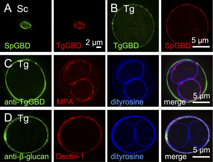
FIG 2 .
Oocyst walls, but not sporocyst walls, of Toxoplasma gondii are labeled with β-1,3-glucan-binding reagents. (A) Walls of Saccharomyces cerevisiae (Sc), deproteinated with NaOH, bind glucan-binding domains from Schizosaccaromyces pombe (SpGBD in green) and Toxoplasma (TgGBD in red) glucan hydrolases, which have been expressed as MBP (maltose-binding protein) fusion proteins and labeled with Alexa Fluor dyes. (B) SpGBD and TgGBD also bind to Toxoplasma gondii (Tg) oocyst walls, but not to sporocyst walls. MBP controls fail to bind to yeast or parasite walls. (C) Antibodies to the Toxoplasma glucan-binding domain (anti-TgGBD in green) bind to oocyst, but not sporocysts, walls of frozen and thawed Toxoplasma. For a control for permeability of sporulated oocysts, the plant lectin MPA (red) that binds to GalNAc labels both the oocyst and sporocyst walls, which are autofluorescent in the UV channel due to the presence of dityrosines. (D) Toxoplasma oocyst walls also are labeled by antibodies to β-1,3-glucan (green) and by the macrophage lectin Dectin-1 (red). See Fig. S2 in the supplemental materialfor fluorescent micrographs of Eimeria oocysts labeled with the same glucan-binding reagents.

