
FIG 3 .
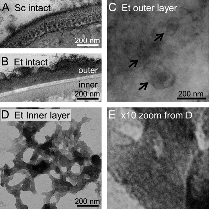
The inner layer of the oocyst wall of Eimeria is a trabecular scaffold. (A) Transmission electron microscopy of cross sections stained with ruthenium red shows that the intact Saccharomyces cerevisiae (Sc) wall is one continuous structure. (B) The Eimeria tenella (Et) oocyst wall is made of an electron-dense outer layer, an electron-lucent interlayer, and a moderately electron-dense inner layer. (C) Negative staining of intact oocysts with ruthenium red shows that the outer layer of the Eimeria oocyst wall is relatively smooth with occasional oval objects that vary in size (arrows). (D) The inner layer of the Eimeria oocyst wall, which is revealed by negatively staining walls that have been mechanically disrupted, is a trabecular scaffold. (E) High-power view of panel D shows putative fibrils of β-1,3-glucan, which are straight and present in parallel arrays, within the trabecular scaffolds. The inner layer of oocyst walls of Toxoplasma are also composed of similar trabecular scaffolds.