FIG 5 .
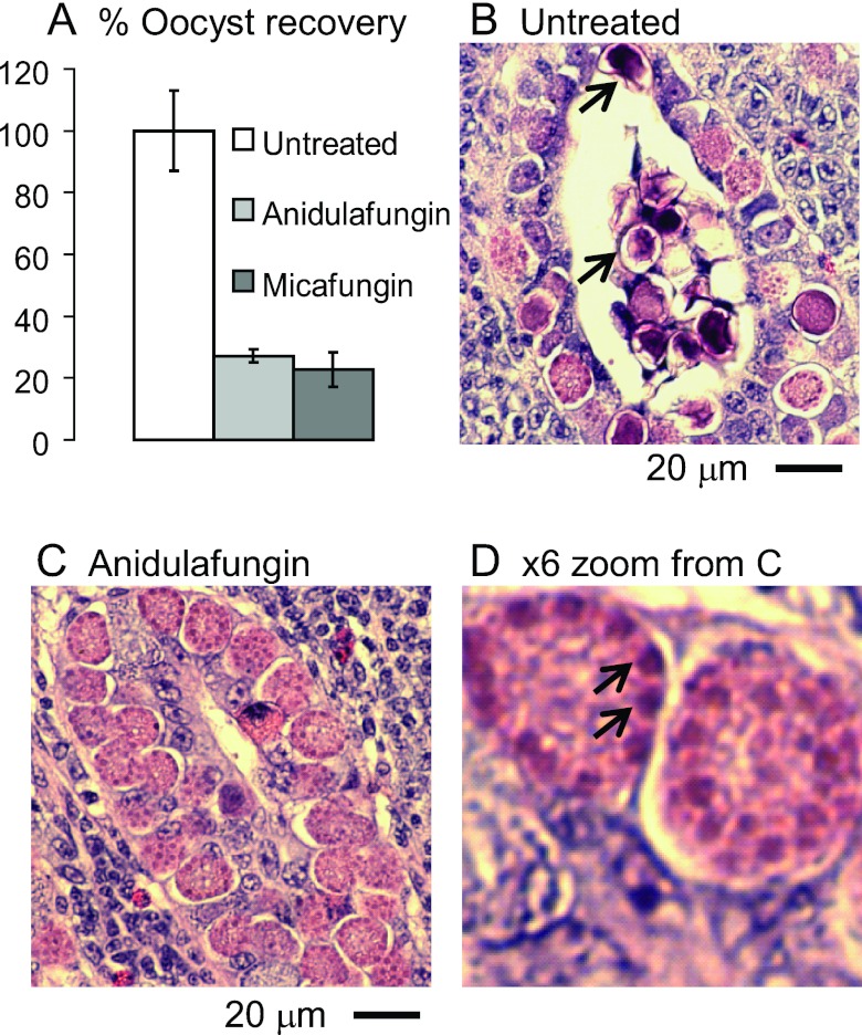
Echinocandins arrest the development of oocyst walls of Eimeria in the ceca of infected chickens. (A) Oocyst recoveries are markedly reduced versus untreated chickens in chickens treated with 10 mg/kg anidulafungin or 10 mg/kg micafungin. Error bars show standard deviations of the oocyst counts in two experiments in which there were two chickens per group. (B) Hematoxylin-and-eosin-stained section of the cecum of an untreated chicken infected with Eimeria shows zygotes in numerous developmental stages, including some with small secretory vesicles (early), large secretory vesicles near the periphery (later), and darkly stained walls (fully developed and often released into the lumen of the crypt) (arrows). (C) A cecum from a chicken treated with anidulafungin shows numerous zygotes that are arrested, so few walled forms are present. Similar results were observed with micafungin. (D) High-power view of panel C shows large purple secretory vesicles at the periphery of developing oocysts arrested by anidulafungin (arrows).

