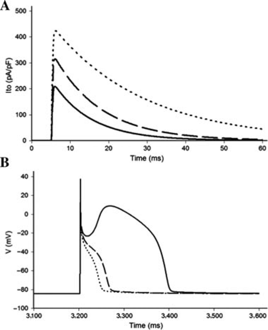
Figure 4.
Effects of p.Val392Ile and p.Gly600Arg on the right ventricular epicardial action potential. A: Simulated Ito current traces during step depolarization for 100 ms to 0 mV from a holding potential of −90 mV. Only first 60 ms are shown. Solid line, wild-type (WT); dashed line, p.Gly600Arg; dotted line, p.Val392Ile. B: Right ventricular epicardial action potentials simulated using WT, p.Gly600Arg, and p.Val392Ile mutant Ito incorporated into a modified Luo–Rudy II AP model. BCL = 800 ms (75 bpm). Solid line, WT; dashed line, p.Gly600Arg; dotted line, p.Val392Ile. Only the fifth AP in the equilibration chain is displayed here, the AP shape does not change in subsequent cycles.

