
Figure 1.
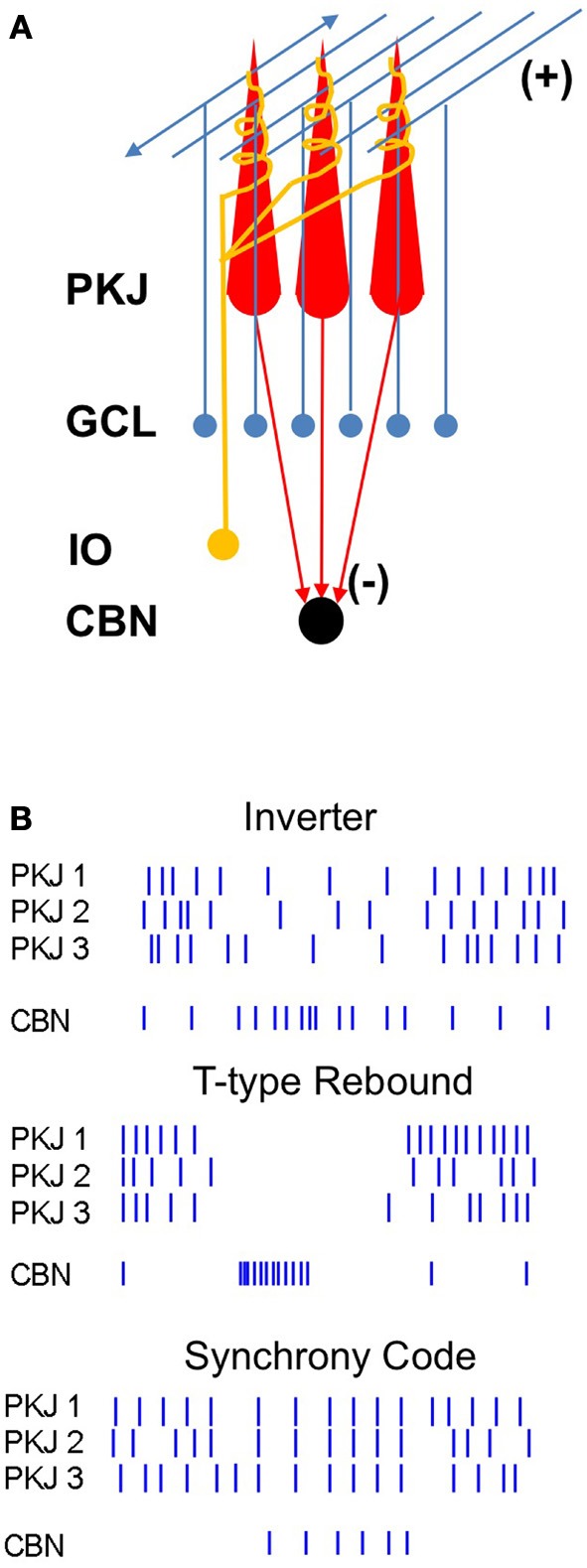
Diagram of the corticonuclear circuit and mechanisms of corticonuclear signaling. (A) Cerebellar Purkinje cells (PKJ) receive glutamatergic inputs from the granule cell layer (GCL) whose axons form ascending inputs onto Purkinje cells and also ramify to form the parallel fibers, as well as from the inferior olive (IO), whose axons form the climbing fibers. Molecular layer inhibitory neurons are not shown. Purkinje cells form GABAergic synapses onto neurons of the cerebellar nuclei (CBN). (B) Schematized spike rasters showing the key features of three non-mutually-exclusive models of Purkinje cell regulation of nuclear cell firing. Nuclear cell spikes reflect responses to three afferent Purkinje cells. Inverter, nuclear cell firing rate varies inversely with Purkinje cell firing rate. T-type Rebound, nuclear cells are largely silenced by Purkinje cell activity, but fire bursts of action potentials driven by low-voltage-activated Ca currents when Purkinje cells stop firing. Synchrony code, nuclear cells are silenced by asynchronous inhibition but produce short-latency spikes after IPSPs from synchronous inputs.