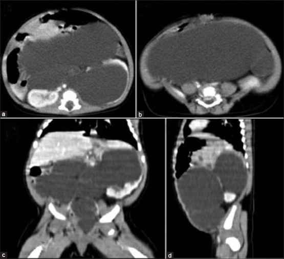
Figure 1.

Post-contrast axial images of computed tomography abdomen and pelvis. (a) and (b) show a large cystic mass filling the entire abdomen and pelvis and arising from the left kidney. (c) Reformatted coronal image and (d) sagittal image further confirms the origin of the mass from upper portion of the left kidney. Inferiorly, the mass is tapering and ending into the lower portion of left side of pelvis
