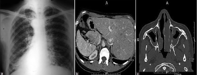
Figure 3.
(a) Cystic bronchiectactic changes in the lower and mid zones with dextrocardia; (b) axial CT image abdomen showing situs inversus with the liver and IVC on the left and the spleen and aorta on the right; (c) axial CT image paranasal sinuses showing mucosal thickening and opacified sinus cavities

