Figure 1.

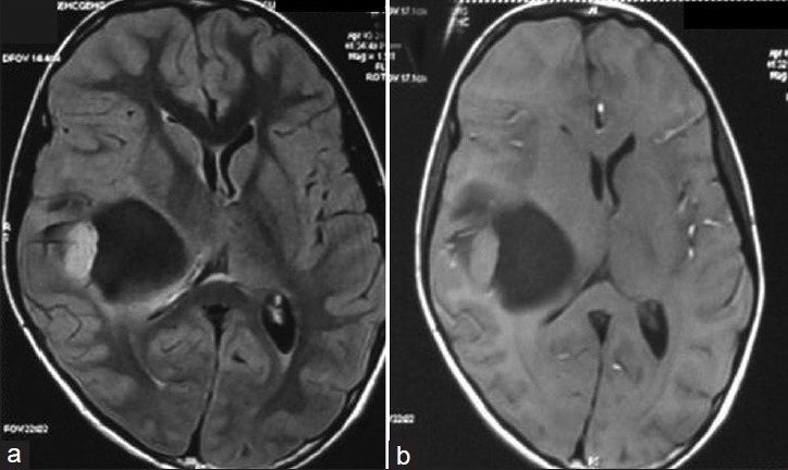
(a and b) FLAIR and T1 contrast MR images of the patient showing a cyst with enhancing mural nodule in the right posterior frontal region. Note that there is no communication to the ventricle

(a and b) FLAIR and T1 contrast MR images of the patient showing a cyst with enhancing mural nodule in the right posterior frontal region. Note that there is no communication to the ventricle