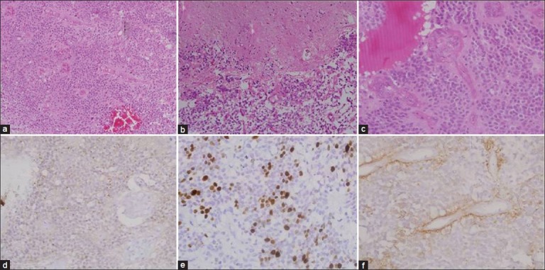
Figure 2.

(a—f) Photomicrograph showing a highly cellular tumor with perivascular arrangement (a) (H and E ×40). The tumor cells have clear cytoplasm and irregularly clumped chromatin with areas of necrosis (b) and endothelial proliferation (c) (H and E ×200). Immunohistochemistry for EMA showing dot positivity (d), MIB-1 indices of 25% (e), and GFAP negativity (f). Overall histological features are suggestive of anaplastic ependymoma with clear cell change
