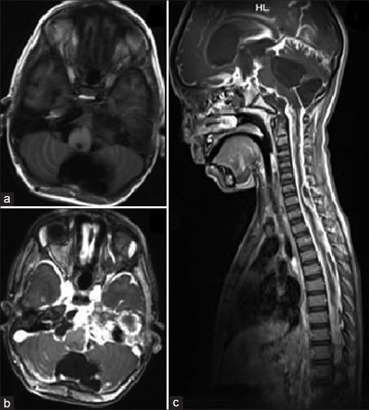
Figure 3.

(a) T1-weighted axial MRI showing minimal lesion in petrous following surgical decompression and chemotherapy. (b) Contrast axial MRI showing enhancement of the leptomeninges. (c) Contrast sagittal MRI showing diffuse enhancement of the leptomeninges of the entire craniospinal axis. The enhancement is so diffuse that it appears like a T2 image
