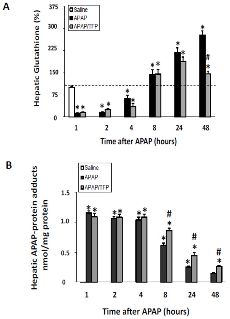
Fig. 4.

Hepatic glutathione and APAP protein adducts in time course study of TFP in APAP toxicity in the mouse. Mice were treated with TFP (10 mg/kg by oral gavage) 1 h prior to APAP (200 mg/kg IP) and sacrificed at the designated times. A. Hepatic glutathione (GSH) levels in APAP and APAP/TFP mice were comparable at 1, 2, 4, 8, and 24 h (*p<0.05) compared to saline, while hepatic GSH was higher in the APAP/TFP mice at 48 compared to the APAP mice at 48 h. B. APAP protein adducts in liver in APAP and APAP/TFP mice compared to saline mice APAP protein adducts were increased to comparable levels in the two APAP groups compared to saline at 1, 2, and 4 h (*p<0.05). Adducts were higher at 8, 24, and 48 h in the APAP/TFP mice compared to APAP mice (#p<0.05).
