
Fig 5.
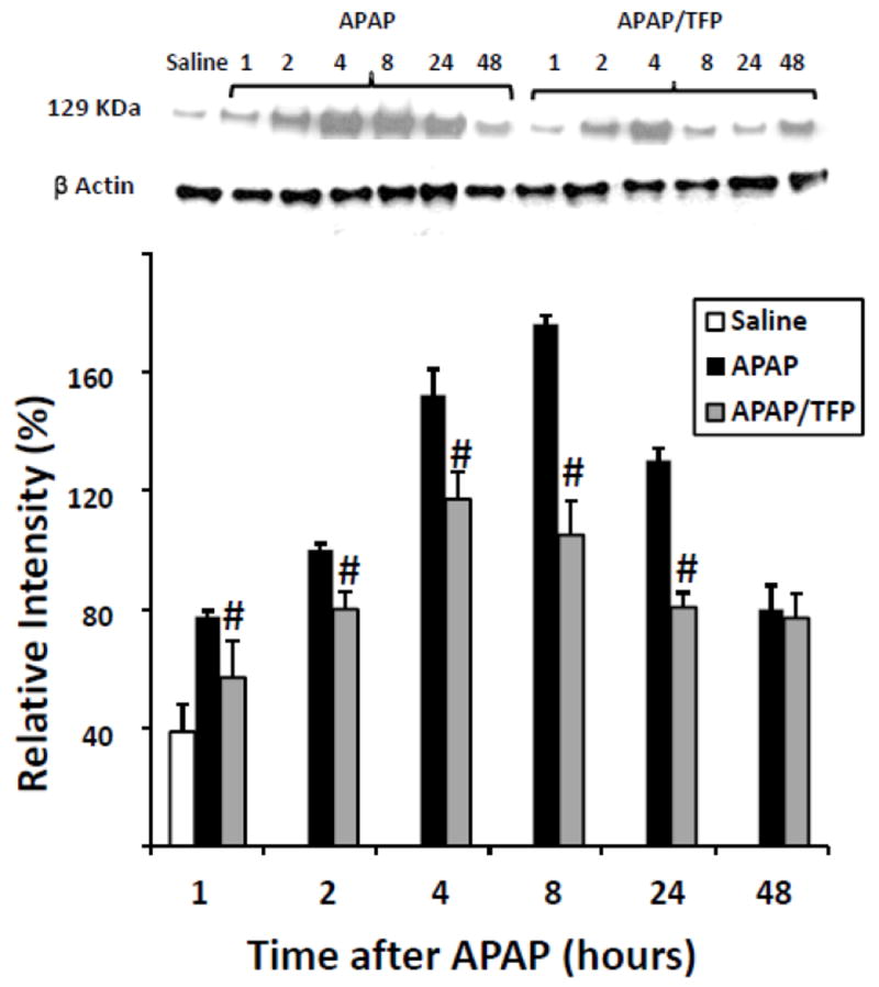
Expression of HIF-1α by immunoblot in APAP and APAP/TFP mice. Nuclear expression of HIF-1α was increased at 1 h in the APAP mice and throughout the time course of APAP toxicity. HIF-1α expression was reduced in the APAP/TFP mice (#p<0.05, compared to APAP mice) through the 24 h time point, as indicated by the immunoblot and confirmed by the densitometry. Representative individual mice are shown in the immunoblot.