
Figure 3.
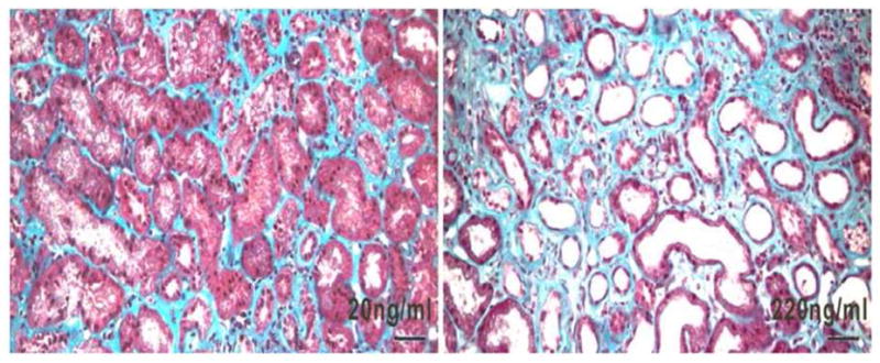
Illustrative kidney biopsies of two patients with membranous nephropathy. Urinary NGAL levels are indicated. Note the widened interstitial compartments, tubular flattening and tubular dilation in the more advanced case which expresses a ten fold higher level of NGAL. Mason trichrome stain. Bar=50μm.