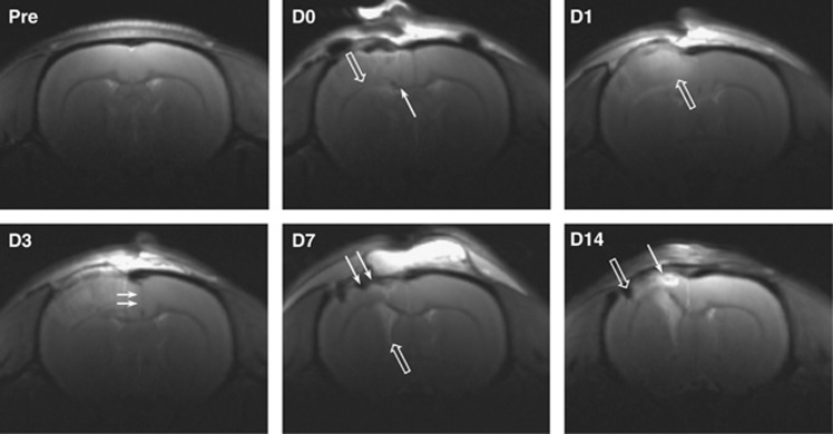
Figure 2.
T2-weighted magnetic resonance imaging (MRI) of a rat brain after controlled cortical impact (CCI). Representative coronal images (bregma −0.5 mm) show the development of the cortical contusion from Day 0 (D0, 1 hour after injury) to Day 14 (D14). Tissue disruption was visible early after injury, with ventral shift of the corpus callosum (open arrow, D0) and frequent small intraparenchymal hemorrhages (line arrow, D0). On D1 and D3, ipsilateral cortical edema was visible as a diffuse tissue hyperintensity (open arrow, D1), and brain swelling was indicated by a midline shift (line arrows, D3). By D7 the swelling had subsided, giving way to ipsilateral cortical thinning (line arrows, D7) and ventricular enlargement (open arrow, D7). By D14 a cortical contusion cyst had developed, filled with hyperintense cerebrospinal fluid (line arrow, D14) and/or hypointense blood products (open arrow, D14).

