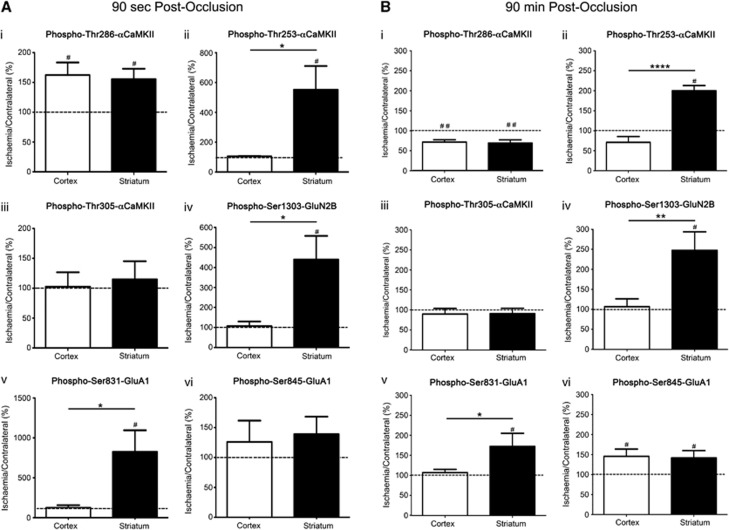
Figure 6.
αCaMKII, GluA1, and GluN2B phosphorylation after occlusion of the middle cerebral artery for 90 seconds or 90 minutes. (i) Thr286-αCaMKII, (ii) Thr253-αCaMKII, (iii) Thr305-αCaMKII, (iv) Ser1303-GluN2B, (v) Ser831-GluA1, and (vi) Ser845-GluA1 phosphorylation in the striatum and cortex of Sprague-Dawley (SD) rats after (A) 90 seconds or (B) 90 minutes occlusion of their middle cerebral artery (n=6/time point). Rats were decapitated, and the striatum and cortex were removed and homogenized. Phosphorylation levels are normalized to total protein expression (i.e., phosphorylation/ total expression), and are expressed as a percentage (ischemia side/contralateral side). #Denotes statistical significance from contralateral side (P<0.05); *denotes statistical significance between the two brain regions (P<0.05). CaMKII, calcium/calmodulin-stimulated protein kinase II.

