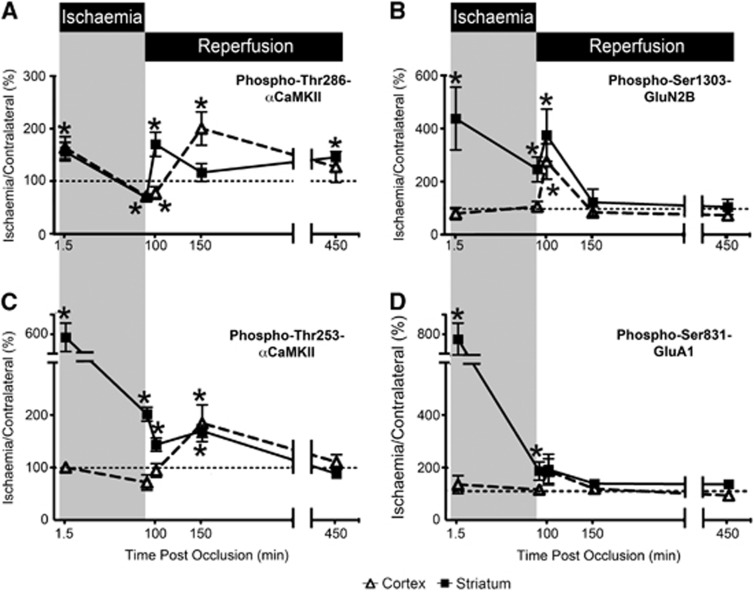
Figure 7.
αCaMKII, GluA1, and GluN2B phosphorylation after varying periods of ischemia and reperfusion. (A) Thr286-αCaMKII, (B) Ser1303-GluN2B, (C) Thr253-αCaMKII, and (D) Ser831-GluA1 phosphorylation in the cortex and striatum of Sprague-Dawley (SD) rats after occlusion of their middle cerebral artery (n=6/time point, except for Ser831-GluA1 and Ser1303-GluN2B 150 and 450 minutes after occlusion where n=3/time point). Rats were decapitated, and the striatum and cortex were removed and homogenized. Phosphorylation levels are normalized to total protein expression (i.e., phosphorylation/total expression), and are expressed as a percentage (ischemia side/contralateral side). Ratios >100% indicate that the level of phosphorylation in the ischemic side is greater than that observed in the contralateral side. Times postocclusion (min) are shown. The data at 1.5 and 90 minutes ischemia are the same as those presented in Figures 7 and 8. *Denotes statistical significance from the contralateral side (P<0.05). CaMKII, calcium/calmodulin-stimulated protein kinase II.

