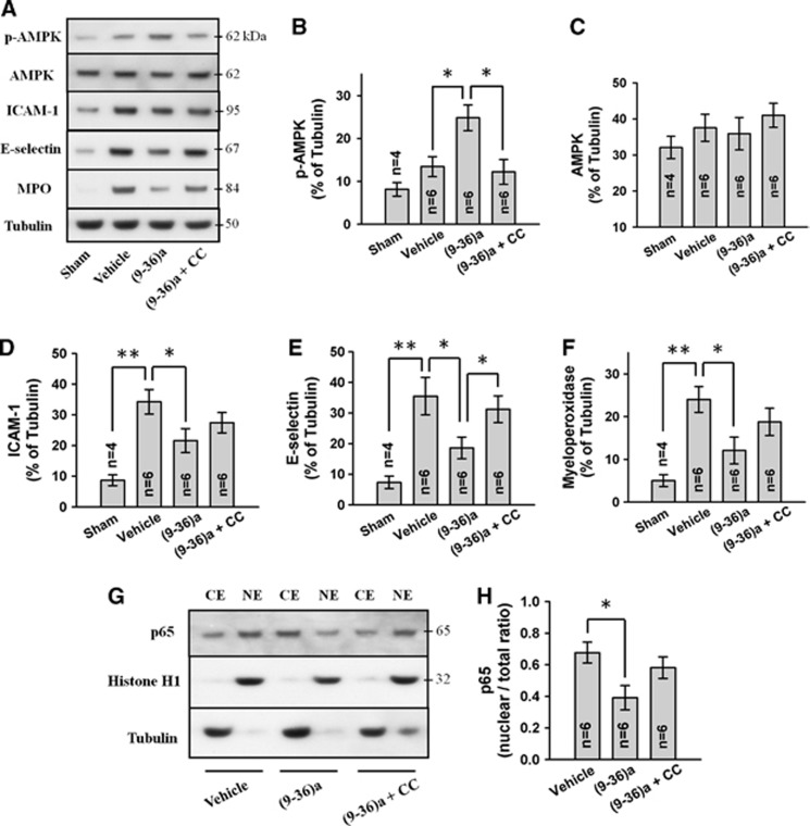
Figure 6.
Representative Western blots (A) of cytoplasmic fraction and effects of GLP-1(9-36)a (1 μg) and compound C (CC; 5 μg) on p-adenosine monophosphate-activated protein kinase (p-AMPK) (B), AMPK (C), ICAM-1 (D), E-selectin (E), and myeloperoxidase (F) levels. Representative Western blots of cytoplasmic (CE) and nuclear (NE) fractions (G) and effects of GLP-1(9-36)a and compound C on NF-κB p65 levels (H). All Western blots were performed on ispilateral cerebral hemisphere at 24 hours after intracerebral hemorrhage (ICH). Expression levels of each protein have been normalized against β-tubulin (CE) or histone H1 (NE). Ex-9=exendin 9-39 (5 μg). GLP-1(9-36)a is abbreviated as (9-36)a. *P<0.05; **P<0.01. GLP, glucagon-like peptide.

