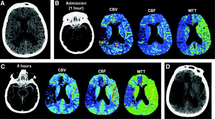
Figure 3.
Absence of reperfusion, even in the setting of complete recanalization, may result in a large follow-up infarct volume. (A) Axial noncontrast computed tomography (CT) carried out on admission (2.5 hours after onset of symptoms) shows subtle hypoattenuation of the right putamen but no sulcal effacement in the right middle cerebral artery (MCA) territory. (B) Axial maximum-intensity projection image from computed tomography angiography (CTA) carried out admission shows occlusion of the M1 segment of the right MCA (arrow). Perfusion CT (PCT) maps show decreased cerebral blood volume (CBV) and cerebral blood flow (CBF) in the right frontal and temporal lobes and a larger region of prolonged mean transit time (MTT) that also involves the right anterior cerebral artery (ACA) territory. The region of decreased CBV corresponds to the infarct core, whereas the surrounding mismatch region of prolonged MTT represents the ischemic penumbra. The patient received endovascular thrombectomy with a MERCI device. (C) Axial maximum-intensity projection image from CTA and PCT carried out 6 hours after admission show that, despite complete recanalization of the right MCA, PCT maps show that the region of decreased CBV and CBF has expanded to include the right anterior ACA territory that was previously considered tissue at risk. MTT is still abnormally increased in the right superficial MCA territory and in a portion of the right ACA territory. (D) Axial noncontrast CT carried out 48 hours after admission shows marked hypoattenuation and edema in the territories matching the perfusion deficit on the reperfusion PCT. Reproduced from Soares et al21 with permission.

