Figure 1.

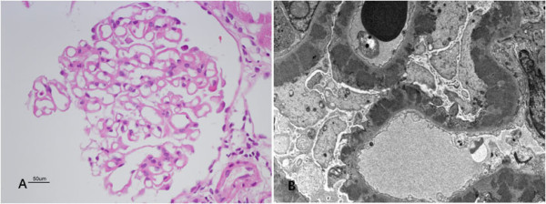
Percutaneous renal biopsy. (A) On light microscopy, glomeruli show a diffuse thickening of basement membrane with normocellularity (Magnification: x400). (B) On electron microscopy, diffuse subepithelial electron dense deposits are easily identified. (Magnification: x2500).
