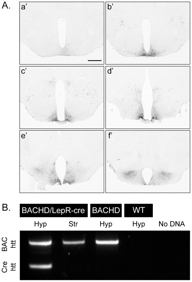
Figure 1. Demonstration and validation of Cre-excision.

(A) GFP staining of hypothalamic sections from crossbred LepR-cre mice×ROSA-eYFP mice illustrates the cre-excision pattern in the hypothalamus, a’f’ represents rostral (bregma −1) to caudal (bregma −2.5) [51] hypothalamic sections, scale bar = 500 µm. (B) PCR analysis confirmed successful excision of human mutant htt exon1 in leptin receptor-expressing neurons in the hypothalamus, but not in a region (striatum), which lacks leptin receptor-expressing neurons.
