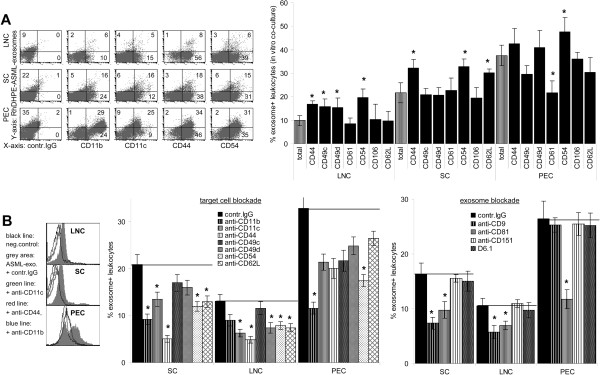
Figure 3.
Adhesion molecules engaged in tumor-exosome uptake. (A) Cells as in (Figure 2A) were stained with adhesion molecule-specific antibodies: representative examples and mean percent ± SD of marker+exosome+ / marker+ cells (3 experiments). (B) LNC, SC and PEC or exosomes were pre-incubated with the indicated antibodies (30 min, 4°C). After washing, cells were co-incubated with dye-labeled exosomes 2h, 4°C: representative examples and mean percent ± SD (3 experiments) of exosome+ cells. (A) Significant differences in the % exosome+marker+ cells as compared to exosome+ cells in the total organ: *, (B) significant differences compared to control IgG treatment: *. There is evidence for an engagement of CD11b, CD11c, CD44, CD49d, CD54 and CD62L in exosome uptake.

