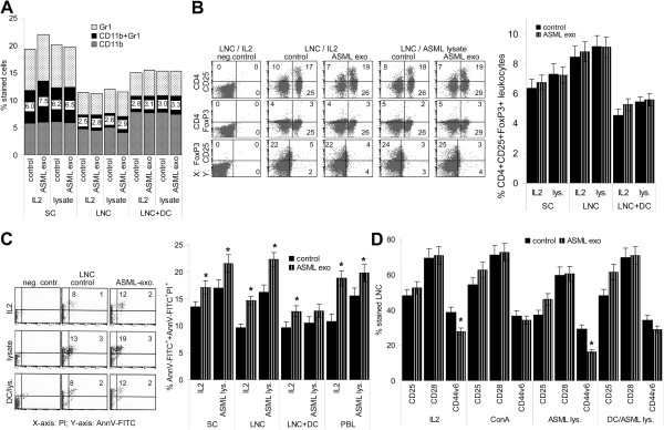
Figure 5.
ASML-exosomes, immunosuppression, apoptosis and activation markers. Leukocytes were stimulated as described in Figure 4. (A) Mean percent (3 experiments) of Gr1+, CD11b+ and Gr1+CD11b+ (MDSC) cells. (B) examples of CD4+CD25+, CD4+FoxP3+ and CD25+FoxP3+ cells and mean percent ± SD (3 experiments) of CD4+CD25+FoxP3+ cells. (C) Representative examples of AnnexinV/PI staining and mean percent ± SD (3 experiments) of AnnV-FITC+/AnnV-FITC+/PI+ cells. (D) Mean percent ± SD (3 experiments) of CD25+, CD28+ and CD44v6+ cells. (C,D) Significant differences in the presence of ASML-exosomes: *. There is no evidence for ASML-exosomes affecting MDSC or Treg. However, apoptosis susceptibility is slightly increased and expansion of CD44v6+ cells is impaired.

