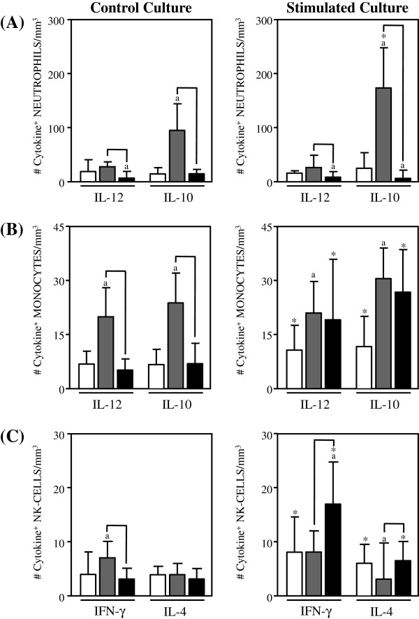
Figure 1.
Analysis of intracytoplasmic cytokine profile of neutrophils. (A), monocytes (B) and NK-cells in the peripheral blood from non treated Indeterminate chagasic patients (IND gray rectangle), Bz-treated Indeterminate chagasic patients (INDt black rectangle) as compared to non-infected individual (NI empty rectangle). Data are expressed as median absolute counts plus the interquartile range of cytokine+ cells observed after short-term in vitro “Control Culture (left panels) and T. cruzi epimastigote antigen “Stimulated Culture (right panels) of whole-blood samples. Cytokine+ neutrophils, monocytes and NK-cells were quantified by dual color immunophenotyping (anti-CD14-TC or anti-CD16-TC plus anti-cytokine-PE mAbs) using specific gate strategies to select each leucocyte subset, as described in Material and Methods. The significant differences at p<0.05 for comparisons with NI are indicated by the letter ‘a. The significant differences at p<0.05 for comparisons with INDt are indicated by connecting lines. The significant differences at p<0.05 for comparative analysis between “Control and “Stimulated cultures are indicated by *.

