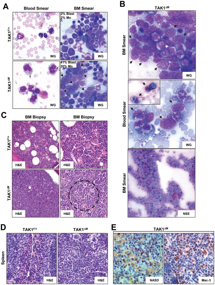
Figure 6. Diseased TAK1ΔM mice developed myelomonocytic leukemia.
(A) Peripheral blood smears (left panels) and bone marrow smears (right panels) from 24-week-old TAK1F/+ and TAK1ΔM mice were stained with Wright-Giemsa (WG) and H&E, respectively. Note numerous monocytes, some with azurophilic granules (arrows) in the peripheral blood and increased blasts in bone marrow aspirate smears of TAK1ΔM mice. Also shown for the BM smear is the percentage of a manual count of blasts and monocytes (Mo). (B) Blood and bone marrow smears from diseased TAK1ΔM mice were stained with Wright-Giemsa (WG) and non-specific esterase (NSE). Note the increased number of blasts in the peripheral blood film and sheets of blasts in the bone marrow aspirate smear (arrows). A subset of blasts was positive for NSE (inset and lower panel). (C) Bone marrow biopsies from 24-week-old TAK1F/+ and TAK1ΔM mice were stained with H&E. Note the increased number of immature cells (dotted circle) in the TAK1ΔM mice. Magnification: left panels, 200x; right panels, 400x. (D–E) Tissue sections of spleens from 24-week-old TAK1F/+ and TAK1ΔM mice were stained with H&E (D) and NASD (E, left panel) and immunostained with an anti-Mac-3 antibody (E, right panel).

