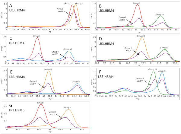
Figure 4.
Variant-specific plasmid DNA duplex infections. Comparison of the different duplex infections possible between variant groups I, II, III and VI. Derivative HRM curves (dF/dT) obtained using primer pairs LR3.HRM4 (A-F) and LR3.HRM6 (G) in real-time PCR HRM assays using variant specific plasmid DNA. All mixed infections shown are the 1:1 duplex artificial mix compared to melting curves generated from single variant reactions.

