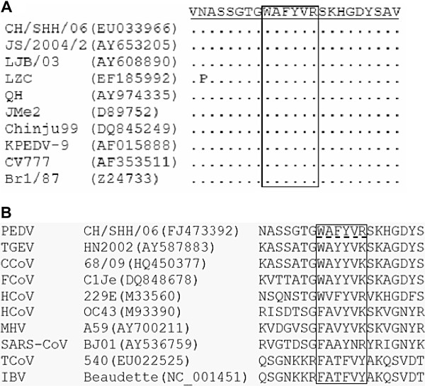
Figure 5.
Alignment of the amino acid sequences of the defined epitope and surrounding region with those of nine PEDV reference strains (A) and nine other coronaviruses (B). The dots represent residues that match the epitope exactly. The homologous regions of different coronaviruses that correspond to the identified epitope are in the box. Abbreviations of each virus and its strain are listed, and the corresponding GenBank accession numbers are shown in parentheses.

