
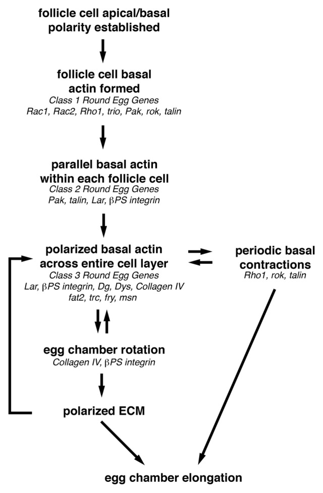
Figure 5. Overview of how egg chamber elongation is achieved. Before egg chamber elongation can begin, a uniformly polarized network of follicle cell basal actin filaments must be established. This requires that the follicle cell basal domain be specified so that the basal actin filaments can be formed, then organized into parallel bundles within each follicle cell that then become oriented perpendicular to the A/P axis of the egg chamber. This uniformly polarized follicle cell basal actin network is then used to provide the force necessary to drive the active migration of the follicle cells over the stationary basal lamina during egg chamber rotation from stage 5–8. As the follicle cells migrate, they organize the ECM components of the basal lamina into parallel arrays that reinforce the orientation of the polarized basal actin filaments and form the molecular corset that contributes to egg chamber elongation. Then during stages 9 and 10, the polarized follicle cell basal actin filaments, together with non-muscle Myosin II, mediate the periodic basal contractions that may maintain the basal actin filaments and contribute to egg chamber elongation by augmenting the molecular corset around the center of the egg chamber. Genes that may function at each step of egg chamber elongation based on their loss-of-function phenotype are indicated in italics.