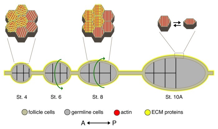
Figure 7. Model of egg chamber elongation. During oogenesis, egg chambers undergo a dramatic change in shape from a sphere (St. 4) to an ellipse (St. 10A). This is achieved through global rotation of the egg chamber within a static basal lamina (St. 5–8) and periodic contractions of a polarized basal actomyosin network (St. 9–10). Diagram depicts a cross section of egg chambers along with a high magnification view of the follicle cell basal surface for a subset of stages. Prior to the onset of egg chamber rotation, the follicle cell basal actin filaments (red) are parallel within each cell but are not uniformly oriented across the follicle cell layer and the ECM protein components of the follicle cell basal lamina (yellow) are unorganized. As the egg chamber rotates (green arrows indicate direction of rotation), the follicle cell basal actin filaments and the ECM fibers become aligned perpendicular to the A/P axis of the egg chamber forming the molecular corset. After the egg chamber stops rotating, a subset of follicle cells, concentrated around the widest point of the egg chamber, undergo periodic contractions of the polarized basal actin filaments, which are mediated by non-muscle Myosin II. These contractions result in a temporary change in the follicle cell basal surface and the generation of an inward force that reinforces the broader molecular corset. St., stage. Adapted from reference 88,and reprinted with permission from Elsevier.
