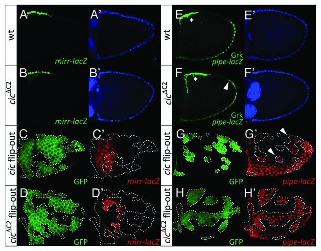
Figure 2. EGFR signaling modulates Cic-mediated repression of mirr. (A and B) Lateral views of stage-10 wild-type (A and A’) and cicΔC2 (B and B’) egg chambers carrying the mirrP2 (mirr-lacZ; ref. 33) enhancer trap. Double stainings with anti-β-galactosidase (A and B, green) and DAPI (A’ and B’, blue) are shown. (C and D) Dorsal views of stage-10 mosaic egg chambers expressing Cic (C and C’) and CicΔC2 (D and D’) proteins in clones marked by expression of GFP (C and D, green). (C’ and D’) show anti-β-galactosidase stainings of mirr-lacZ expression (red). Only CicΔC2 causes full repression of mirr-lacZ. (E and F) Lateral views of stage-10 wild-type (E and E’) and cicΔC2 (F and F’) egg chambers carrying the M2-lacZ (pipe-lacZ; ref. 12) reporter. Stainings with anti-β-galactosidase and anti-Gurken antibodies (E and F, green) and DAPI (E’ and F’, blue) are shown; the Gurken signals are indicated with asterisks. A single genomic cicΔC2 transgene (see ref. 22) leads to expanded pipe-lacZ expression by an average of 1.3 cells (n = 20) in the dorsal-posterior region (arrowhead in panel F). (G and H) Lateral views of stage-10 mosaic egg chambers expressing Cic (G and G’) and CicΔC2 (H and H’) proteins in clones marked by expression of GFP (G and H, green). G’ and H’ show anti-β-galactosidase stainings of M1-lacZ (pipe-lacZ; ref. 12) expression (red). Note that Cic-expressing clones show partial derepression of pipe-lacZ close to the endogenous pipe domain (arrowheads, G’), whereas CicΔC2 causes robust derepression of the reporter in all positions.

An official website of the United States government
Here's how you know
Official websites use .gov
A
.gov website belongs to an official
government organization in the United States.
Secure .gov websites use HTTPS
A lock (
) or https:// means you've safely
connected to the .gov website. Share sensitive
information only on official, secure websites.
