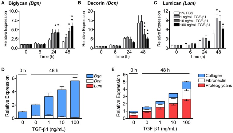
Figure 4. TGF-β1 increased the expression of biglycan and decreased the expression of decorin.
(A–C) The mean expression (± SEM) of proteoglycan genes coding for biglycan, decorin, and lumican was evaluated in tenocyte-seeded collagen gels after treatment with control media or 1–100 ng/mL of TGF-β1 over 48 hours. Biglycan expression was significantly increased in the 10 and 100 ng/mL treatment groups at 24 and 48 hours (p<0.05, Panel A). Decorin expression, on the other hand, increased 14-fold in the control and 1 ng/mL treatment groups, but this increase was suppressed by 10 and 100 ng/mL of TGF-β1 (p<0.05, Panel B). Lumican expression was significantly increased with 1 and 10 ng/mL TGF-β1 (p = 0.001), but not 100 ng/mL TGF-β1 (Panel C). N = 5−6 gels per treatment per time point. *p<0.05 vs. control media, •p<0.05 vs. 1 ng/mL TGF-β1. (D) Inter-gene analysis of the proteoglycans revealed that biglycan expression was the highest at all treatments and time points (82–98%), followed by decorin (2–17%) and lumican (<2%). (E) The relative transcript levels of the proteoglycans, fibronectin, and collagen were also determined. At each treatment and time point, proteoglycans (red) were expressed most highly, followed by fibronectin (white) and collagen (blue). All categories of ECM genes were upregulated in a dose-dependent manner by 1–100 ng/mL of TGF-β1 at 48 hours.

