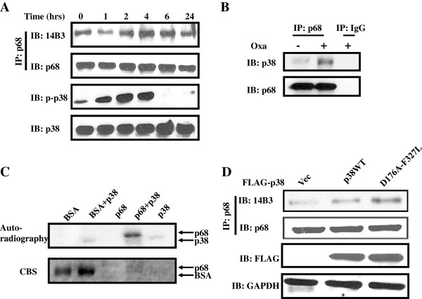
Figure 2.
MAPKPhosphorylation of p68 by p38 MAPK. (A) Threonine phosphorylations of p68 in HCT116 cells that are treated with 20 μM of oxaliplatin for different times are analyzed by immunobloting the p68 that are immunoiprecipitated (IP:p68) from cell lysates using antibody against phorsphor-threonine (IB:14B3). Phosphorylation of p38 MAPK under the same treatment is analyzed by immunoblot of cell lysates using antibody against the phosphorylated p38. Immunoblot of p68 (IB:p68) in the immunoprecipitates indicate the amounts of p68 that are precipitated. Immunoblot of p38 in the cell lysate (IB:p38) indicate the cellular levels of p38, as a loading control. (B) Co-immunoprecipitation of p38 and p68 in the cell extracts of HCT116 cells with/without oxaliplatin treatment (Oxa, +/− 20 μM) was analyzed by immunoblot of p68 immunoprecipitates (IP:p68) using antibody against p38 (IB:p38). Immunoblot of p68 (IB:p68) in the immunoprecipitates indicate the amounts of p68 that are precipitated. IP:IgG is the immunoprecipitation using rabbit IgG, serving as a negative control IP. (C) Phosphorylation of recombinant His-p68 or BSA, as a control, by recombinant p38 in the presence of [γ-32P]-ATP is revealed by autoradiography. The amounts of proteins used in the phosphorylation reactions are shown by coomasie blue stains (CBS). (D) Phosphorylation of p68 by exogenous expression of Flag-tagged p38 MAPK, wild type and constitutively active mutant D176A-F327L, in HCT116 cells are analyzed by immunoblotting the p68 that are immunoiprecipitated (IP:p68) from cell lysates using antibody against phorsphor-threonine (IB:14B3). Immunoblot of p68 (IB:p68) in immunoprecipitates indicate the amounts of p68 that are precipitated. Immunoblot of Flag-tag (IB:FLAG) indicate the exogenous p38 levels. Immunoblot of GAPDH (IB:GAPDH) is a loading control.

