Figure 3.
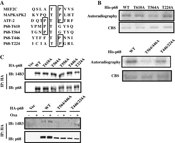
Phosphorylation site(s) of p68 by p38 MAPK. (A) Prediction of potential p38 MAPK phosphorylation site(s) in the p68 reading frame and compared to the consensus p38 MAPK phosphorylation sites of several authentic p38 MAPK substrates by a web-based phosphorylation site prediction program NetPhos 2.0, (B) Phosphorylation of recombinant His-p68 and mutants with single site mutation (Upper) and double site mutations (Lower) by recombinant p38 in the presence of [γ-32P]-ATP is revealed by autoradiography. The amounts of proteins used in the phosphorylation reactions are shown by coomasie blue stains (CBS). (C) Phosphorylation of exogenously expressed HA-p68s, wild type (WT) and mutants (Single site mutation, Upper, and Double site mutations, Lower), in HCT116 cells with/without oxaliplatin treatment (Oxa, +/−) are analyzed by immunoblotting the p68 that is immunoiprecipitated (IP:p68) from cell lysates using antibody against phorsphor-theronine (IB:14B3). Immunoblot of p68 (IB:p68) in immunoprecipitates indicate the amounts of p68 that are precipitated.

