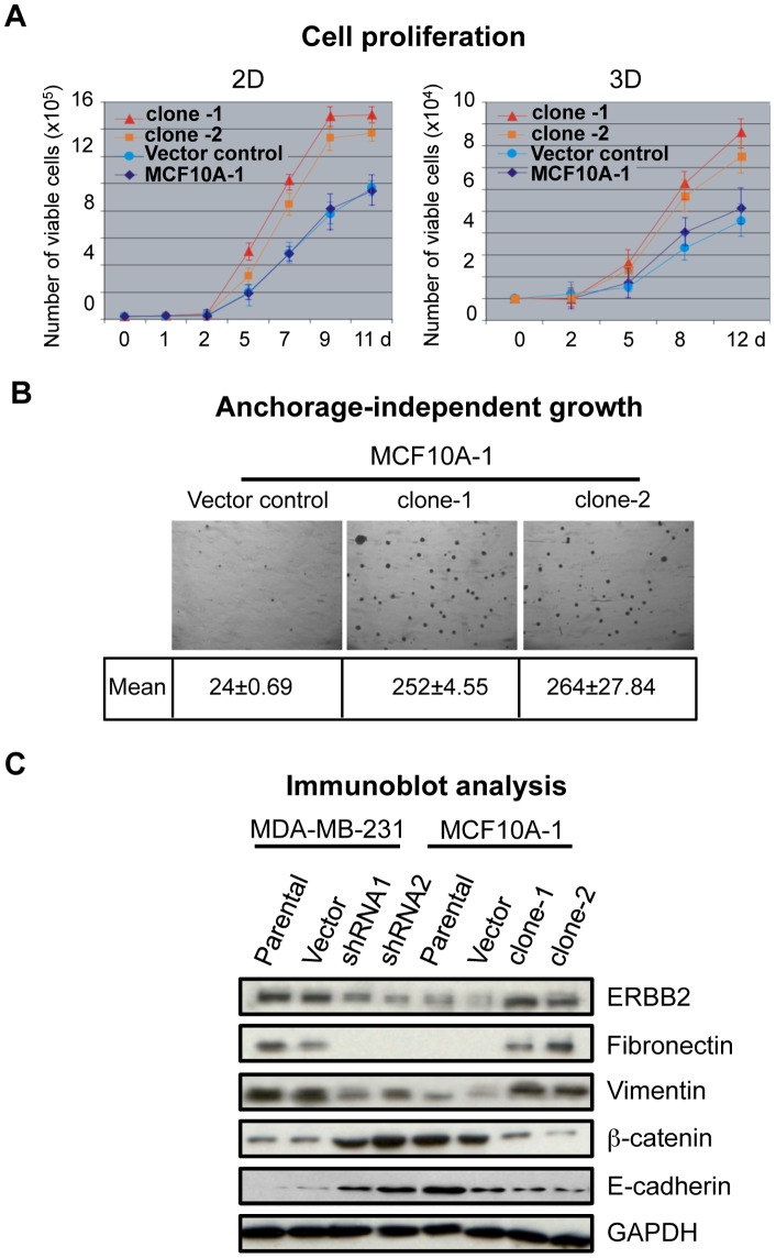
Figure 2. SATB1 overexpression induces EMT in MCF10A-1 cells.
A) Cell proliferation assay to compare the growth over time (days, d) of the parental MCF10A-1 and vector control cells with single-cell-derived SATB1-overexpressing MCF10A-1 clones clone-1 and clone-2 on plastic dishes (2D) or on Matrigel (3D). Error bars indicate ±s.e.m. from three independent experiments. B) Representative photographs of soft agar colonies formed by control and SATB1 overexpressing (clone-1 and clone-2) MCF10A-1 cells after 25 days of culture. The mean colony counts from three replicates are shown. C) Immunoblot analyses for the expression of mesenchymal markers (fibronectin and vimentin), epithelial markers (E-cadherin and ß-catenin) as well as SATB1 target ERBB2 in MDA-MB-231 (parental, control and SATB1-depleted by shRNA1 or shRNA2) and MCF10A-1 (parental, control, clone-1 and clone-2). Cell lysates were prepared from cells cultured on plastic dishes (2D). GAPDH was used as a loading control.

