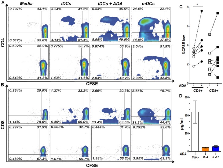
Figure 5. ADA enhances DCs immunogenicity in iDC-T-cell Allogeneic cocultures.
In A and B, iDCs, obtained as described in the Materials and Methods, from a representative healthy donor were cultured during 48 h in medium in the absence of ADA (iDCs), in the presence of 2 µM ADA (iDCs+ADA) or in the presence of maturating cocktail (mDCs). DCs were washed and cocultured with allogeneic (upper contour plots) or autologous (lower contour plots) T-cells (1∶20 DCs:T-cells ratio). After 7 days, the percentage of CD4+ (A) and CD8+ (B) T-cell proliferation was assessed by flow cytometry using the CFSE method. In (C) percentages of CD4+ (circles) and CD8+ (squares) T-cell proliferation in the absence (open symbols) or the presence (filled symbols) of 2 µM ADA in allogeneic cocultures from 6 to 7 healthy donors are shown. Each pair of linked symbols represents results from a particular healthy donor. *P<0.05. In (D) bars indicate IFN-γ, IL-4 and IL-17 levels in ADA-treated iDC co-culture supernatants. Results are the mean ± SD (pg/ml) of 3 independent experiments.

