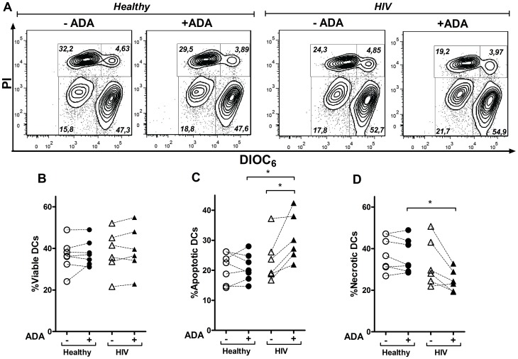
Figure 7. Effect of ADA on the iDCs viability.
iDCs, obtained as described in the Materials and Methods, from healthy or HIV-infected donors were cultured during 48 h in the absence (−ADA) or in the presence (+ADA) of 2 µM ADA. Cell viability was assessed through DIOC6 and propidium iodide (PI) staining and measured by flow cytometry. In (A), contour plots showing the percentage of viable (bright DIOC6 and negative propidium iodide staining), apoptotic (low DIOC6 and negative propidium iodide staining) and necrotic (low DIOC6 and positive propidium iodide staining) populations from a representative healthy or HIV-infected donor are shown. Percentage of viable (B), apoptotic (C) and necrotic (D) DCs from 7 different healthy and 6 HIV-infected donors are shown.*P<0.05.

