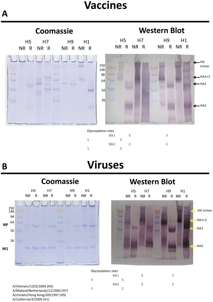
Figure 4. Polyacrylamide gels run under reducing and nonreducing conditions for trimeric HA and HA1 and HA2 subunits bound and separate.
Coomassie blue stain was used for protein and western blots with polyclonal antisera for protein identity. Gels are for vaccines (Figure 4A) and viruses (Figure 4B). Baculovirus expressed H3 HA was used as control (not shown).

