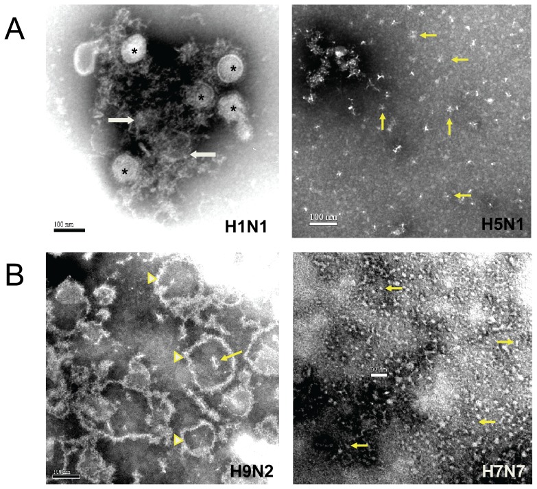
Figure 5. Selected electron micrographs of vaccines illustrating the morphologic structures described in Table 4 .
Figure 5A shows intact and split virus particles (asterisks) in the influenza A/Cal/04 (H1N1) subunit vaccine (CSL) of Table 1 along with structures of indistinct morphology (arrows). Also shown in Figure 5A is an EM of the A/Vietnam/04 (H5N1) subunit vaccine (SP) of references 5, 6, 10 in Table 1 that was selected to show a large number of stellate structures (arrows point to examples) although indistinct structures similar to those in the H1N1 vaccine were dominant in the H5N1 vaccine (not shown). Figure 5B is an EM of the influenza A/CK/G9 (H9N2) vaccine (Novartis) of Table 1 that shows the predominant varying size particles of membrane with external projections as well as a number of stellates, one apparently within an empty particle (arrow). Also in Figure 5B is an EM of the influenza A/Mallard (H7N7) vaccine (SP, ref. 12 in Table 1) that primarily showed small (5 to 20 nm) round and elongated structures (arrows point to examples). Some stellates and a rare intact particle were also seen.

