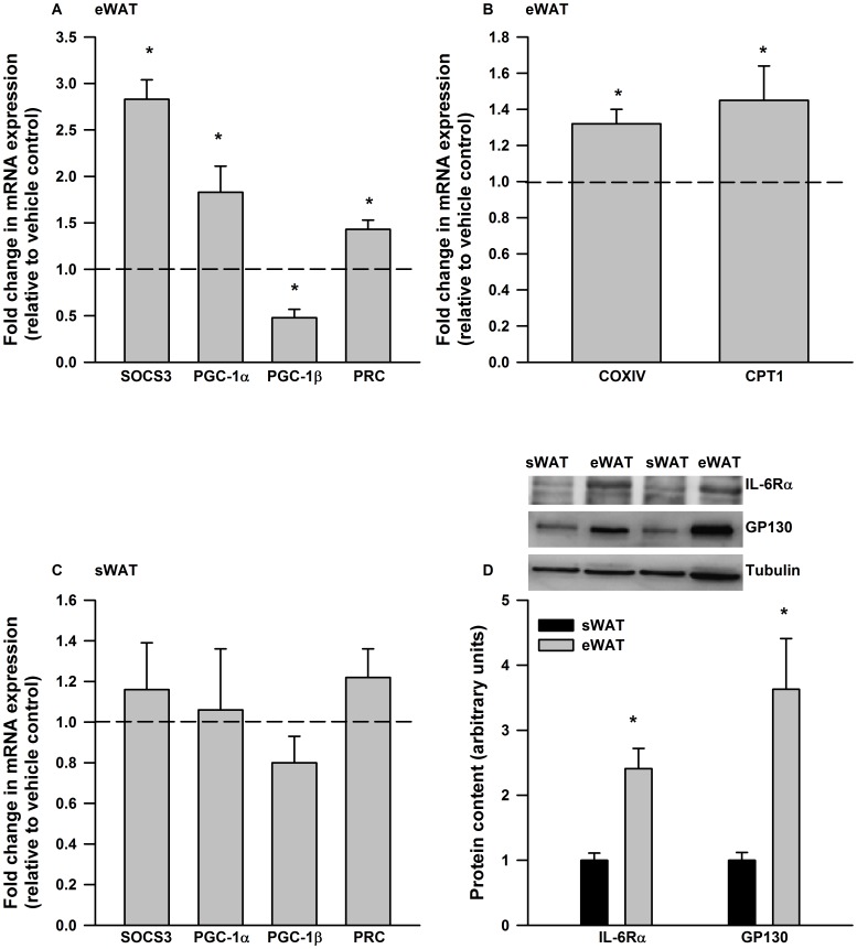
Figure 1. IL-6 exerts depot specific differences in gene expression in mouse adipose tissue.
IL-6 treatment (6 hours, 75 ng/ml) increased the expression of A) SOCS3, PGC-1α and PRC mRNA in cultured epididymal adipose tissue (eWAT). B) A 12 hr IL-6 (75 ng/ml) treatment increased the expression of COXIV and CPT-1 in cultured eWAT. C) Cultured subcutaneous adipose tissue (sWAT) was not responsive to IL-6 treatment (75 ng/ml, 6 hours) and this was associated with D) reductions in the protein content of IL-6 receptor alpha and GP130. Data are presented as means + SE for 7 cultures, each from an individual mouse, per group and is expressed relative to the vehicle treated control culture from the same animal. For the Western blot data in D) data are presented as means + SE for 8–10 mice per group. Representative Western blots are presented above the quantified data. * P<0.05.

