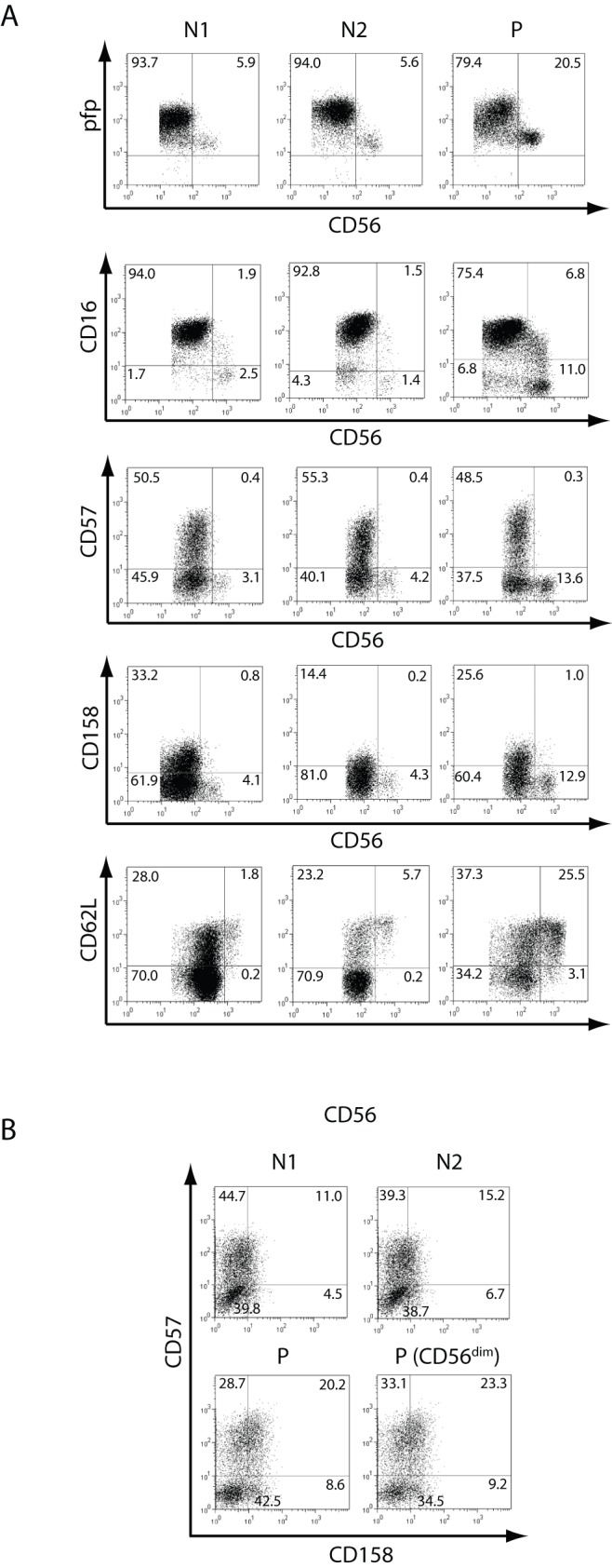
Figure 2. Phenotype of NK cells.

A) PBMCs from two normal (healthy) donors (N1 and N2) and from the patient (P) were stained with anti-CD3 and anti-CD56, and anti-pfp, anti-CD16, anti-CD57, anti-CD158 (KIR) or anti-CD62L, and fluorescence derived from this third fluorochrome-labeled mAb was depicted against CD56 after gating NK cells as CD3−CD56+ as dot plots. B) Expression of CD57 and CD158 in NK cells (depicted as dot plots and gated as CD3−CD56+ cells) from N1, N2 and the patient (P), and on CD3−CD56dim cells from P. The percentage in each quadrant is shown. Results correspond to one from 3 independent experiments. PBMCs from blood samples obtained in May 2007 were used.
