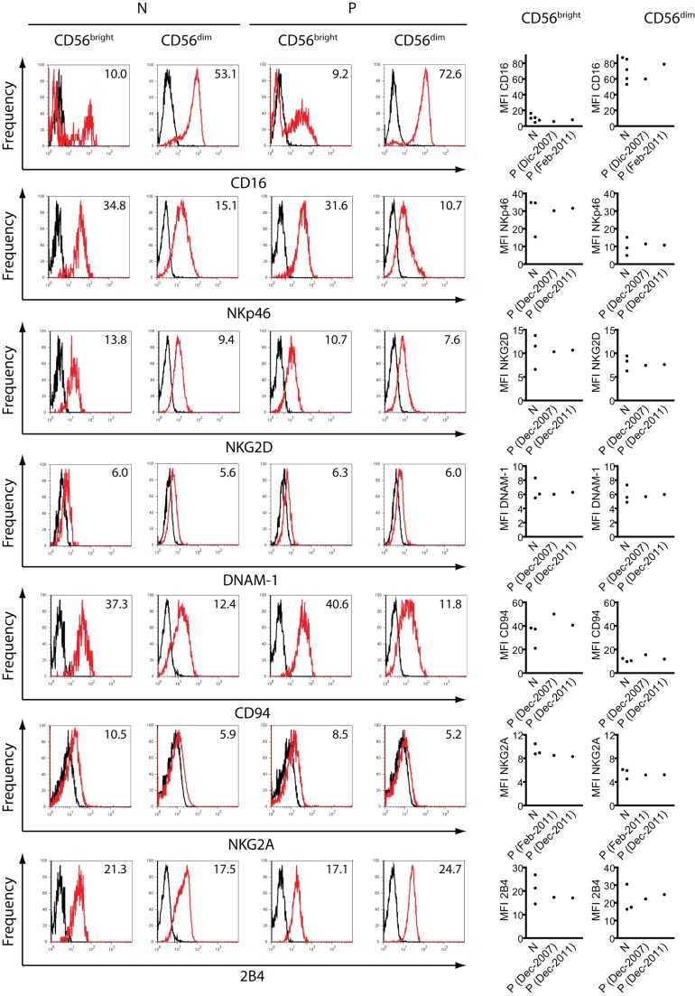
Figure 3. Expression of NK cell receptors.
PBMCs from at least 3 different normal(healthy) donors (N) and from different blood draws from different dates (indicated in parentheses) from the patient (P) were stained with anti-CD3, anti-CD56, and anti-CD16, anti-NKp46, anti-NKG2D, anti-DNAM-1, anti-CD94, anti-NKG2A or anti-2B4 as described in Methods. Fluorescence derived from the third fluorochrome-labeled mAb on CD3−CD56bright or CD3−CD56dim cells was depicted. Representative histograms are shown on the left with the corresponding MFI inserted in the histrogam, while the MFI for each receptor of the healthy normal donors and the 2 different samples of the patient are depicted on the right graphs. PBMCs from blood samples obtained in December 2011 were used.

