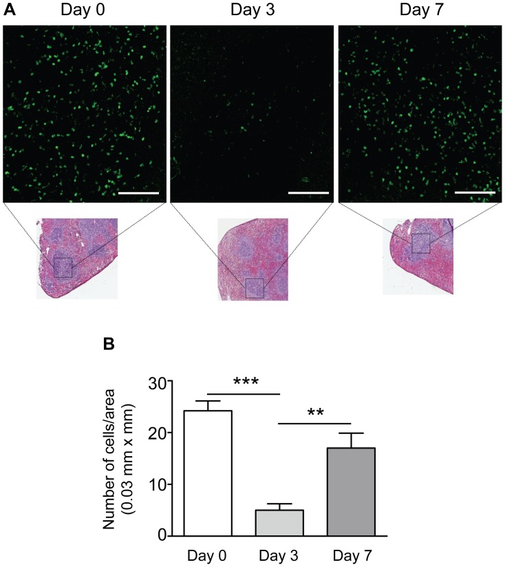
Figure 4. Regulatory T cells emigrate from the spleen after injury of the carotid artery.
A. Representative confocal images showing FoxP3-GFP+ cells (green) in the spleen of non-operated control mice (Day 0) and in the spleens of mice 3 or 7 days after injury. Smaller insets show spleen sections from the same mice stained for H&E for visualization of tissue architecture and the white pulp areas that were imaged. Scale bars = 100 µm. B. Summarized data from confocal experiments showing significantly reduced number of FoxP3-GFP+ cells in spleens 3 days after injury and restored levels at day 7. N = 5 mice per group; ***P < 0.001, **P < 0.01.

